The nervous system is one of the most complex and poorly understood biological systems in the human body. Neurological and psychiatric disorders — including Alzheimer's disease, Parkinson's disease, schizophrenia, autism spectrum disorder, epilepsy, multiple sclerosis, and glioblastoma — affect hundreds of millions of people worldwide and remain among the most challenging conditions to diagnose, understand, and treat. At BioinformaticsNext, we provide expert bioinformatics support for neuroscience and CNS research, helping scientists decode the molecular architecture of the brain and nervous system using the latest sequencing technologies and computational approaches.
Bioinformatics for Neuroscience & CNS Genomics
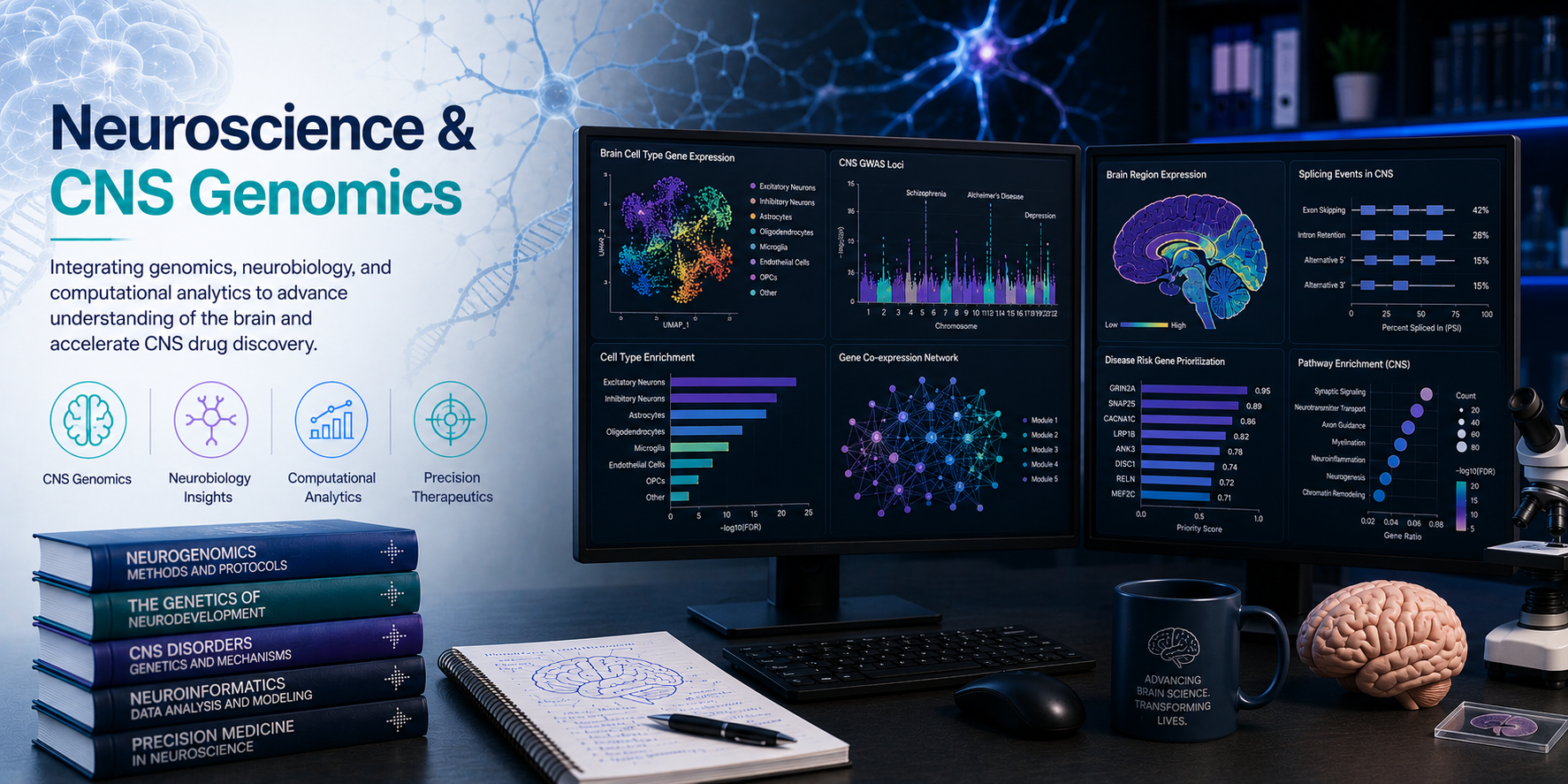
Decoding the molecular architecture of the brain — from single-cell atlases to neurodegeneration genomics.
Advances in single-cell sequencing, spatial transcriptomics, epigenomics, and multi-omics integration are transforming our understanding of the nervous system — enabling the construction of comprehensive cell atlases of the brain, the identification of disease-associated cell types and molecular pathways, and the discovery of novel therapeutic targets for neurological and psychiatric conditions.
Making sense of this data requires specialised bioinformatics expertise tailored to the unique challenges of CNS research: extreme cellular heterogeneity, rare cell populations, region-specific gene expression, and the complex interplay between neurons, glia, and the neural vasculature. At BioinformaticsNext, we provide the computational tools and biological insight to extract meaningful discoveries from your neuroscience data.
What We Analyse
Comprehensive CNS profiling across cell types, brain regions, disease states, and genetic risk.
- Cell-type-specific gene expression across brain regions and developmental stages
- Neuronal, glial, and vascular cell type identification and characterisation
- Disease-associated transcriptomic and epigenomic changes in neurodegeneration
- Genetic risk variants and their functional impact in neurological disorders
- Brain-region-specific chromatin accessibility and regulatory element activity
- Spatial organisation of cell types and gene expression within brain tissue
- RNA splicing dysregulation in ALS, FTD, and other CNS conditions
- Multi-omics integration for CNS biomarker discovery and drug target identification
Our Neuroscience & CNS Genomics Services
Comprehensive bioinformatics support across the full range of neuroscience and CNS genomics data types and research questions.
All pipelines follow published best-practice guidelines and are version-controlled for full reproducibility.
1. Bulk RNA-seq of Brain Tissue & Neural Cells DEG · Deconvolution · Splicing · WGCNA
Bulk RNA-sequencing of brain regions, dissected tissue, or cultured neural cells provides a comprehensive transcriptomic snapshot of gene expression changes in CNS disease models, patient post-mortem tissue, and experimental perturbations.
- Alignment & quantification — STAR 2-pass alignment to human or mouse brain reference transcriptomes; Salmon quasi-mapping for rapid quantification; gene- and transcript-level abundance estimation
- Differential gene expression — DESeq2, edgeR, and limma-voom for identifying genes dysregulated between disease vs. control, brain regions, treatment conditions, or developmental timepoints
- CNS pathway enrichment — GSEA and fGSEA with neurological disease gene sets; synaptic, neuroinflammatory, mitochondrial, and autophagy pathway analysis; GO and KEGG enrichment for CNS-relevant terms
- Co-expression network analysis — WGCNA for brain-region-specific gene module identification; hub gene discovery and module correlation with clinical variables (age, disease stage, cognitive score)
- Cell-type deconvolution from bulk RNA-seq — Estimating neuronal, astrocyte, microglia, oligodendrocyte, and endothelial cell proportions from bulk brain RNA-seq using CibersortX, MuSiC, and DWLS with single-cell reference atlases
- Alternative splicing in CNS — rMATS and SUPPA2 for exon-level splicing changes; cryptic exon detection relevant to ALS (TDP-43, FUS), SMA (SMN2), and other splicing-related neurological disorders
2. Single-Cell RNA Sequencing for Neuroscience scRNA-seq · Allen Atlas · DAM · iPSC · Organoids
The brain contains hundreds of distinct cell types — and single-cell RNA-seq has become the gold standard for characterising this cellular diversity in health and disease. We support the complete scRNA-seq analytical workflow optimised for neural tissue, with experience across human post-mortem brain, mouse brain, iPSC-derived neurons, organoids, and dissociated neural cultures.
- Neural tissue-specific QC — Adapted thresholds for neuronal and glial cell populations; ambient RNA removal with SoupX and DecontX; doublet detection with Scrublet and DoubletFinder; dissociation-induced stress gene filtering
- Cell-type annotation — Automated annotation with SingleR and Azimuth using Allen Brain Atlas and Human Cell Atlas brain references; manual curation with canonical marker genes for excitatory neurons, inhibitory neurons, astrocytes, microglia, oligodendrocytes, OPCs, and endothelial cells
- Neuronal subtype classification — Layer-specific cortical neuron identification (L2/3, L4, L5, L6); interneuron subtype mapping (PV, SST, VIP, LAMP5); hippocampal, cerebellar, and brainstem cell-type characterisation
- Disease cell state identification — Disease-associated microglia (DAM), reactive astrocytes (A1/A2), oligodendrocyte stress states, and neurotoxic cell state profiling in Alzheimer's, Parkinson's, and ALS datasets
- Differential abundance testing — Milo and scCODA for detecting compositional shifts in brain cell populations between disease and control, or across treatment conditions
- Trajectory & pseudotime analysis — Monocle3, scVelo, and Palantir for mapping neuronal differentiation, glial activation trajectories, and disease progression state transitions
- Multi-sample integration — Harmony, scVI, and Seurat RPCA for integrating datasets across donors, brain banks, and sequencing batches; inter-individual variation handling for post-mortem studies
3. Spatial Transcriptomics of Brain Tissue Visium · SPARK-X · cell2location · Allen Atlas
The brain is a spatially organised organ — and spatial transcriptomics preserves the anatomical context of gene expression, enabling the study of region-specific cell distributions, laminar organisation, white matter tract gene expression, and the spatial relationship between cell types in disease.
- 10x Visium brain tissue processing — SpaceRanger alignment to brain reference; spot-level QC and normalisation; brain region annotation using Allen Brain Atlas coordinates
- Spatially variable gene identification — SPARK-X, SpatialDE, and Moran's I for genes with spatially structured expression across brain sections; region-specific marker gene discovery
- Cell-type deconvolution of brain spots — RCTD, SPOTlight, and cell2location for mapping neuronal and glial cell types onto spatial coordinates; layer-specific cell-type distribution analysis
- Brain region boundary delineation — Automated identification of cortical layers, hippocampal subfields, white matter, and grey matter boundaries from spatial gene expression data
- Plaque and pathology-associated spatial analysis — Spatial mapping of amyloid plaque-associated transcriptional changes; identification of peri-plaque reactive cell states in Alzheimer's disease spatial datasets
- scRNA-seq to spatial mapping — Projection of single-cell brain atlases onto spatial coordinates for high-resolution cell-type positioning within tissue architecture
4. CNS Genetics & GWAS Analysis PLINK2 · SuSiE · PsychENCODE · Brain eQTL
Large-scale GWAS studies have identified hundreds of loci associated with neurological and psychiatric disorders — but translating these genetic signals into biological understanding and therapeutic targets requires sophisticated post-GWAS bioinformatics. We provide comprehensive genetic analysis services for CNS research.
- Neurological disorder GWAS analysis — Quality-controlled association analysis for Alzheimer's disease, Parkinson's disease, schizophrenia, bipolar disorder, ASD, ADHD, epilepsy, and multiple sclerosis using PLINK2, REGENIE, and SAIGE
- Post-GWAS fine-mapping — Credible set construction and causal variant prioritisation with SuSiE and FINEMAP; colocalisation with brain eQTL datasets (GTEx Brain, PsychENCODE, CommonMind)
- Brain eQTL integration — Linking CNS GWAS signals to brain-region-specific and cell-type-specific eQTLs from GTEx v8, PsychENCODE, and single-cell eQTL resources
- Rare variant analysis in neurological disorders — Gene-based burden and SKAT tests for rare coding variants; WES and WGS-based rare variant discovery in familial neurological disease
- Polygenic risk score (PRS) development — PRS construction and validation for neurological and psychiatric disorders; PRS association with disease subtype, severity, and treatment response
- Mendelian randomisation for CNS targets — Two-sample MR using brain-expressed gene variants as instruments to assess causal effects on neurological disease outcomes; drug target validation
5. Epigenomics of the Brain & Nervous System ATAC · ChIP · EWAS · scATAC · Methylation
The epigenome plays a central role in neural development, neuronal identity, synaptic plasticity, and the long-term consequences of environmental exposures on brain function. Dysregulation of epigenetic mechanisms is increasingly implicated in neurological and psychiatric disorders.
- Brain ATAC-seq analysis — Open chromatin profiling in neurons, glia, and iPSC-derived neural cells; cell-type-specific accessible chromatin identification; neurological disease-associated chromatin accessibility changes
- ChIP-seq in neural tissue — Histone modification mapping (H3K27ac, H3K4me3, H3K27me3) in brain tissue and neural cell types; active enhancer and bivalent promoter identification; TF binding site mapping
- Brain DNA methylation analysis — EPIC array and WGBS analysis of post-mortem brain tissue; DMR identification in Alzheimer's, Parkinson's, and psychiatric disorders; epigenetic clock analysis for brain ageing
- Neurological EWAS — Epigenome-wide association studies for neurological and psychiatric traits with cell-type composition correction; methylation QTL mapping in brain tissue
- Single-cell epigenomics of neural cells — scATAC-seq analysis with ArchR and SnapATAC2; cell-type-specific chromatin accessibility in the brain; 10x Multiome joint RNA + ATAC analysis in neural tissue
6. Neurodegeneration & Disease Genomics Alzheimer's · Parkinson's · ALS · FTD · Neuroinflammation
Neurodegenerative diseases — including Alzheimer's disease, Parkinson's disease, ALS, FTD, Huntington's disease, and the prion diseases — are driven by a complex interplay of genetic risk, protein aggregation, neuroinflammation, and progressive neuronal loss. We provide targeted bioinformatics support for neurodegeneration research across all major disease areas.
- Alzheimer's disease multi-omics — Integration of GWAS, brain RNA-seq, proteomics, and methylation data; APP, PSEN1/2, APOE, and TREM2 pathway analysis; amyloid and tau pathway dysregulation profiling
- Parkinson's disease genomics — SNCA, LRRK2, GBA, and PINK1/Parkin pathway analysis; mitochondrial dysfunction and autophagy gene expression profiling; substantia nigra transcriptomics
- ALS / FTD analysis — TDP-43, FUS, C9orf72, SOD1 variant analysis; cryptic exon splicing detection; UPS and autophagy pathway dysregulation; motor neuron-specific transcriptomics
- Neuroinflammation profiling — Microglial activation state characterisation; complement pathway, cytokine, and chemokine expression analysis; TREM2, CD33, and CLU neuroinflammation gene analysis
- Protein aggregation pathway analysis — Proteostasis, heat shock response, and UPS gene expression profiling; aggregation-prone protein interaction network analysis
- Brain ageing & resilience genomics — Transcriptomic and epigenomic changes across the ageing brain; resilience vs. vulnerability gene signatures; longevity gene expression analysis
7. iPSC & Brain Organoid Transcriptomics iPSC · Organoids · CRISPR Screens · Isogenic
Induced pluripotent stem cell (iPSC)-derived neurons, glia, and brain organoids are transforming neuroscience research — enabling disease modelling in human cells with patient-specific genetic backgrounds. We provide specialised bioinformatics support for iPSC and organoid transcriptomics studies.
- iPSC differentiation transcriptomics — Bulk and single-cell RNA-seq analysis of neuronal differentiation protocols; maturation state assessment; comparison with primary neural cell references
- Brain organoid scRNA-seq — Cell-type composition analysis of cerebral, cortical, hippocampal, and midbrain organoids; developmental trajectory inference; batch correction across organoid lines
- Disease vs. isogenic control comparison — Differential expression between patient-derived and gene-corrected isogenic iPSC lines; variant-specific transcriptomic effects
- Neuronal maturity & subtype scoring — Transcriptomic maturity scoring relative to primary human fetal and adult references; excitatory vs. inhibitory neuron subtype proportion quantification
- CRISPR screen analysis in neural cells — MAGeCK and BAGEL2-based genome-wide CRISPR screen analysis in iPSC-derived neurons; essential gene and disease modifier identification
Key Applications
Research and translational applications across neuroscience and CNS disease.
- Single-cell brain atlas construction and annotation
- Alzheimer's and Parkinson's disease transcriptomics
- Neurological disorder GWAS and post-GWAS analysis
- Brain region and layer-specific gene expression profiling
- Neuroinflammation and microglial activation characterisation
- iPSC-derived neuron and organoid transcriptomics
- ALS, FTD, and splicing disorder RNA analysis
- Psychiatric disorder multi-omics (schizophrenia, bipolar, ASD)
- Brain epigenomics and DNA methylation in disease
- CNS drug target identification and validation
- Spatial transcriptomics of brain tissue sections
- Brain ageing, resilience, and longevity genomics
Our Analytical Workflow
A structured, reproducible process from initial data assessment to final interpreted results and written report.
Step 1 — Project Scoping Free
We discuss your brain region, cell type, disease model, data type, and research questions to define the most appropriate analytical approach and deliverables.
Step 2 — Data Receipt & QC
Secure encrypted data transfer; comprehensive QC (FastQC, MultiQC, Cell Ranger summary) with neural tissue-specific thresholds before analysis begins.
Step 3 — Pipeline Configuration
Version-controlled pipeline setup with Snakemake or Nextflow; reference selection (human GRCh38, mouse GRCm39, or custom assemblies) and tool configuration matched to your assay and CNS data type.
Step 4 — Primary Analysis
Alignment, cell demultiplexing, variant calling, or methylation extraction as appropriate; all intermediate files retained for full auditability.
Step 5 — Cell-Type & Disease Analysis
Neural cell-type annotation, disease state profiling, differential abundance, or pathway analysis depending on your research question.
Step 6 — Statistical Analysis
Differential expression, enrichment analysis, GWAS association, or epigenomic differential analysis with appropriate correction for multiple testing and CNS-specific confounders.
Step 7 — Visualisation
Publication-ready figures — UMAP embeddings with cell-type labels, brain region heatmaps, spatial transcriptomics maps, volcano plots, pathway diagrams, and genome browser tracks.
Step 8 — Report & Manuscript Support Optional
Full written report with methods, results, and biological interpretation; optional manuscript methods section and figure legend preparation.
Tools & Technologies
Validated, peer-reviewed, and actively maintained tools across all neuroscience and CNS genomics pipelines.
- scRNA-seq: Seurat, Scanpy, Cell Ranger, SingleR, Azimuth
- Neural Annotation: Allen Brain Atlas references, Human Cell Atlas brain
- Trajectory: Monocle3, scVelo, Palantir, PAGA
- Integration: Harmony, scVI, Seurat RPCA, BBKNN
- Bulk RNA-seq: STAR, DESeq2, edgeR, WGCNA, rMATS
- Deconvolution: CibersortX, MuSiC, DWLS, SCDC
- Spatial: SpaceRanger, Squidpy, RCTD, cell2location
- GWAS & Genetics: PLINK2, REGENIE, SAIGE, SuSiE, coloc
- Epigenomics: MACS3, bismark, ChAMP, ArchR, chromVAR
- CRISPR Screens: MAGeCK, BAGEL2, CRISPRcleanR
- Pathway Analysis: clusterProfiler, GSEA, fGSEA, ReactomePA
- Visualisation: ggplot2, ComplexHeatmap, dittoSeq, Seaborn
- Multi-Omics: MOFA+, MixOmics, SNF
- Workflow: Snakemake, Nextflow, CWL
Reference Databases & Resources We Use
All major neuroscience and CNS genomics reference resources for comprehensive biological annotation and interpretation.
- Allen Brain Atlas — Comprehensive brain-region-specific gene expression reference for human and mouse brain; cell-type marker gene databases for neural annotation
- Human Cell Atlas — Brain — Single-cell reference atlases of the human brain for cell-type annotation across regions and developmental stages
- PsychENCODE — Brain-specific eQTL, chromatin accessibility, and gene expression data for psychiatric and neurological disorder analysis
- CommonMind Consortium — Post-mortem brain RNA-seq and genomics data for schizophrenia, bipolar disorder, and ASD
- GTEx v8 — Brain Regions — Tissue-specific eQTL data across 13 brain regions for CNS GWAS signal functional annotation
- ROSMAP / MSBB / Mayo RNAseq — Alzheimer's disease post-mortem brain omics datasets from Religious Orders Study and Rush Memory and Aging Project
- UK Biobank / iPSYCH — Large-scale neurological and psychiatric GWAS cohorts for PRS development and genetic architecture analysis
- DisGeNET — Gene-disease association database for neurological and psychiatric disorder gene prioritisation
- SynGO — Synaptic gene ontology database for synaptic function and plasticity gene enrichment analysis
Project Deliverables
A complete, structured set of outputs ready for publication, grant submission, or translational application.
- Quality control report (MultiQC HTML + PDF) for all samples
- Processed data files: aligned BAMs, count matrices, annotated cell-type labels
- Differential expression tables, pathway reports, and cell-type composition summaries
- Publication-ready figures (PDF, SVG, PNG at 300 dpi)
- Full written report: methods, results, biological interpretation, and recommendations
- Pipeline scripts and configuration files for full reproducibility
- Post-delivery consultation call for results walkthrough and Q&A
- Interactive R Shiny brain atlas explorer application
- Manuscript methods section and figure legends (journal-formatted)
- Supplementary data tables and extended figure sets
- Allen Brain Atlas-aligned spatial visualisation outputs
- Training and knowledge transfer sessions for your team
- Long-term retainer support for ongoing neuroscience projects
Why Choose BioinformaticsNext?
Deep neurobiological expertise combined with validated, scalable computational pipelines — delivering results that are biologically meaningful, statistically rigorous, and publication-ready.
CNS Domain Expertise
Our analysts understand the unique biology of the brain and nervous system — from neuronal cell-type diversity to neurodegeneration pathways — ensuring your data is interpreted in its full biological context.
Neural Tissue Specialisation
We apply CNS-specific QC thresholds, reference datasets, and annotation resources — not generic single-cell workflows — for every neuroscience project.
End-to-End Service
From raw sequencing files to annotated brain cell atlases and written reports — every step handled in-house with no need for your own bioinformatics infrastructure.
Fast Turnaround
Most projects are delivered within 2–4 weeks. Rush turnarounds are available for grant deadlines and conference submissions.
Multi-Omics Integration
We integrate scRNA-seq, spatial transcriptomics, epigenomics, genetics, and proteomics data from brain tissue — providing a comprehensive molecular view that no single assay can deliver alone.
Flexible Engagement
Project-based, hourly, or long-term retainer arrangements tailored to your timeline and budget with no minimum commitment.
Data Security
Encrypted data transfer and storage. NDAs and GDPR-compliant Data Processing Agreements available upon request.
Global Reach
UK-headquartered with clients across Europe, North America, the Middle East, and Asia-Pacific.
Frequently Asked Questions
Common questions from neuroscience and CNS genomics clients.
Yes. Post-mortem brain tissue presents specific challenges including RNA degradation (reflected in RIN scores), post-mortem interval effects, and cellular composition variability between donors. We apply appropriate QC filters, RIN-aware normalisation, and cell-type deconvolution strategies specifically designed for post-mortem brain datasets. We have experience with data from the Allen Brain Bank, ROSMAP, Mayo RNAseq, and other major brain banking initiatives.
Yes. We regularly work with iPSC-derived neuron, astrocyte, and microglia transcriptomic data, as well as cerebral and cortical brain organoid scRNA-seq datasets. We use primary human fetal and adult brain references for maturity assessment and cell-type benchmarking, and can compare your iPSC-derived cells directly against in vivo references.
Brain tissue scRNA-seq is among the most challenging single-cell analysis contexts due to the vast diversity of neuronal and glial subtypes. We use brain-specific reference datasets (Allen Brain Atlas, Human Cell Atlas Brain), multi-resolution clustering, and iterative sub-clustering to resolve rare and closely related cell populations. We also apply dissociation stress gene filtering to remove transcriptional artefacts introduced during tissue dissociation.
Yes. We regularly integrate newly generated datasets with public brain omics data from GTEx, PsychENCODE, Allen Brain Atlas, ROSMAP, CommonMind Consortium, and the Human Cell Atlas — enabling comparative analysis across brain regions, disease states, and donors that would not be possible from a single dataset alone.
Absolutely. We assist with the bioinformatics and genomics sections of neuroscience grant applications — including study design, proposed analytical workflows, power calculations, and preliminary data generation. Please get in touch as early as possible in the grant preparation process.
Related Research Areas & Services
Neuroscience and CNS genomics intersects with multiple other research domains we support.
- Genetics & Genomics — Neurological disorder GWAS, rare variant analysis, Mendelian randomisation, and polygenic risk score development for CNS diseases
- Cancer & Oncogenomics — Brain tumour genomics including glioblastoma multiforme (GBM), medulloblastoma, and CNS metastasis profiling
- Immunology & Immuno-Oncology — Neuroinflammation, microglial biology, and neuroimmunology — the immune-brain interface in neurodegeneration and CNS tumours
- Structural & Functional Genomics — Brain epigenomics, chromatin accessibility in neural cells, and regulatory element characterisation in CNS development and disease
- Drug Development & AI Discovery — CNS drug target identification, blood-brain barrier penetration prediction, and neurological disease biomarker development
Ready to Advance Your Neuroscience Research?
Tell us about your brain tissue, your sequencing data, and your research questions. Our neuroscience and CNS genomics team will design a tailored analytical plan — typically within 48 hours of your enquiry. Whether you are building a single-cell brain atlas, profiling post-mortem tissue from neurodegeneration patients, or identifying CNS drug targets from GWAS data, we are here to support you from day one.